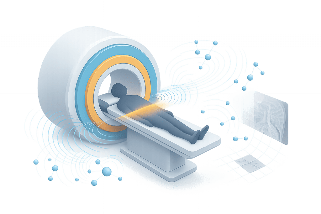
Magnetic resonance imaging (MRI) turns the tiny magnetic moments of hydrogen nuclei into detailed pictures of soft tissues. In a strong magnetic field, nuclei precess at the Larmor frequency; radio-frequency pulses tip them, gradients encode position, and a Fourier transform reconstructs images whose contrast depends on T1, T2, and sequence parameters.
Table of Contents
- From Nuclear Spin to Signal: The Core Idea
- Building an MRI System
- Encoding Space: Slice, Frequency, and Phase
- Contrast and Parameters: T1, T2, Proton Density and Beyond
- From Raw Data to Images: Reconstruction and Quality
1) From Nuclear Spin to Signal: The Core Idea
MRI works because many biological tissues are rich in hydrogen (water and lipids). Each hydrogen nucleus (a single proton) behaves like a tiny bar magnet with spin. When a patient enters a powerful static magnetic field (B₀), these spins align slightly with the field and precess at a characteristic rate known as the Larmor frequency (proportional to B₀). This sets the stage for generating measurable signals.
Excitation. An applied radio-frequency (RF) pulse at the Larmor frequency deposits energy and tips the net magnetization away from the B₀ axis. The amount of tip (the flip angle) depends on the pulse strength and duration. Immediately after the pulse, the spins are synchronized (in phase) and induce a small voltage in a receive coil—a decaying oscillation called the free induction decay (FID).
Relaxation. Once the RF energy is removed, two recovery processes begin:
- T1 (longitudinal) relaxation: the magnetization returns toward alignment with B₀ as energy is exchanged with the surrounding lattice.
- T2 (transverse) relaxation: the in-phase coherence between spins decays due to local magnetic interactions, causing the measured signal to fade.
Importantly, T1 and T2 vary by tissue, which is why tissues can be made brighter or darker by choosing specific sequence timings. Brain white matter, gray matter, muscle, fat, edema, and lesions often separate cleanly in at least one contrast setting. The art of MRI is choosing parameters that accentuate useful differences while minimizing scan time and safety.
Why hydrogen? Hydrogen is abundant and has a strong magnetic moment, yielding higher signal-to-noise ratio (SNR) than rarer nuclei. Other nuclei (¹³C, ²³Na, ³¹P) are possible in research settings but produce weaker signals and longer scan times.
An MRI scanner is a carefully choreographed orchestra of fields and electronics designed to create, disturb, and listen to nuclear magnetization with exquisite precision.
2) Building an MRI System
Main components.
- Main magnet (B₀): Provides a strong, uniform static field—commonly 1.5T or 3T in clinical systems. Stronger B₀ boosts SNR and spectral separation but can increase artifacts and safety constraints.
- Gradient coils (Gx, Gy, Gz): Superimpose linearly varying fields along x, y, and z to encode position. Fast, powerful gradients enable thin slices and high resolution but can raise acoustic noise and peripheral nerve stimulation risk.
- RF chain: Includes transmit coils that deliver tuned pulses and receive coils (often multi-channel arrays) that pick up the faint signals. Coil geometry strongly impacts SNR and penetration depth.
- Control and reconstruction computer: Orchestrates pulse sequences, timing, data sampling, and converts k-space data into images.
To minimize lists and emphasize relationships, the table below summarizes how each block contributes to image formation:
| Subsystem | Primary Role | Key Trade-off it Influences |
|---|---|---|
| Main Magnet (B₀) | Sets Larmor frequency, boosts SNR | Higher field improves SNR but can increase susceptibility artifacts and impose stricter safety limits |
| Gradient Coils | Spatial encoding for slice selection, frequency and phase | Faster gradients → higher resolution and faster imaging, but more noise and stimulation risk |
| RF Transmit | Excites spins (flip angle control) | Larger flip angles can improve contrast but elevate SAR (specific absorption rate) |
| RF Receive | Detects decaying signal (SNR) | More elements closer to anatomy improve SNR and parallel imaging performance |
| Recon Computer | Sampling, Fourier transform, corrections | Handles speed/quality trade-offs, parallel imaging, and artifact suppression |
Safety note. MRI uses non-ionizing radiation, which is a key advantage. However, safety management is still critical: SAR (RF heating), projectile risks from ferromagnetic objects, and implants compatibility must be controlled through screening and protocols.
3) Encoding Space: Slice, Frequency, and Phase
Turning a global magnetic resonance into a spatial map requires precise position encoding. MRI does this by modulating the Larmor frequency with gradients so that frequency and phase correlate with location.
Slice selection. To excite a single slice, an RF pulse is played with a gradient (e.g., along z). Only spins whose Larmor frequency falls within the RF pulse bandwidth get tipped—defining a thin slice. Slice thickness depends on RF bandwidth and gradient strength; thinner slices improve anatomical detail but reduce SNR.
Frequency and phase encoding. In-plane encoding typically uses:
- A frequency-encoding gradient (readout) during signal acquisition so that each x-position resonates at a distinct frequency.
- A phase-encoding gradient applied briefly before readout along the perpendicular axis (y), imparting position-dependent phase shifts.
Collecting data across many phase-encoding steps fills k-space, a frequency-domain representation of the image. The center of k-space governs overall contrast and SNR, while the outer regions encode fine detail. The final image emerges from a Fourier transform of this k-space matrix.
Accelerations. Modern systems shorten scan time by skipping portions of k-space or sampling more efficiently, then recovering missing information computationally:
- Partial Fourier and parallel imaging (e.g., SENSE/GRAPPA) exploit coil sensitivity differences to reduce phase-encoding lines.
- Compressed sensing uses sparsity to reconstruct acceptable images from randomized undersampling, especially in angiography or dynamic studies.
Artifacts and remedies. Because encoding is delicate, motion can smear phase information (ghosting), metal can disturb local fields (susceptibility), and insufficient sampling can alias distant structures into the field of view. Practical mitigation includes faster sequences, breath-holds, motion correction, metal-artifact-reduction techniques, and careful parameter selection.
4) Contrast and Parameters: T1, T2, Proton Density and Beyond
Image appearance depends on how long we wait before listening and how we refocus the spins. Three timing knobs dominate:
- TR (repetition time): delay between excitations; controls degree of T1 recovery.
- TE (echo time): time from excitation to signal readout; longer TE allows more T2 decay to influence contrast.
- TI (inversion time): used in inversion-recovery sequences to null a chosen tissue.
T1-weighted imaging. Short TR and relatively short TE make tissues with shorter T1 (like fat) appear brighter and those with longer T1 (like CSF) darker. T1 weighting highlights anatomy and post-contrast enhancement patterns.
T2-weighted imaging. Longer TE accentuates differences in transverse coherence; tissues with longer T2 (often fluid) appear bright. This is valuable for detecting edema and many pathologies that increase water content.
Proton density (PD) weighting. Using a long TR and short TE minimizes T1/T2 effects so brightness reflects the number of mobile protons. PD images emphasize tissue cellularity and subtle structure with reduced contrast among tissues that share similar proton densities.
Spin-echo vs gradient-echo.
- Spin-echo (SE) sequences employ a 180° refocusing pulse that reverses dephasing from static inhomogeneities, producing clean T2 contrast.
- Gradient-echo (GRE) sequences omit the 180° pulse, are faster, and are sensitive to T2* (which includes field inhomogeneity effects). GRE is useful for dynamic imaging, angiography, and detecting blood products or calcifications.
Inversion recovery and special contrasts.
- FLAIR (fluid-attenuated inversion recovery) sets TI to null CSF, revealing periventricular lesions.
- STIR (short TI inversion recovery) suppresses fat, often used in musculoskeletal imaging where fat suppression aids lesion conspicuity.
- Diffusion-weighted imaging (DWI) sensitizes the sequence to microscopic water motion; restricted diffusion appears bright, helpful in acute stroke and cellular tumors.
- fMRI (functional MRI) typically uses BOLD contrast—tiny oxygenation-related changes in T2*—to map neural activation.
To keep things concrete yet compact, the table below summarizes the most common weightings:
| Contrast Type | What It Highlights | Typical Knobs |
|---|---|---|
| T1-weighted (SE/GRE) | Anatomy, fat, post-contrast enhancement | Short TR, short TE; optional flip-angle tweaks in GRE |
| T2-weighted (SE) | Fluid, edema, inflammation | Long TR, long TE |
| Proton Density (SE) | Proton concentration with minimal T1/T2 bias | Long TR, short TE |
| FLAIR (IR) | Lesions near CSF by suppressing free fluid | TI tuned to null CSF, moderate TR/TE |
| DWI | Restricted diffusion (e.g., acute ischemia) | b-value selection; echo-planar readout |
| T2*-weighted GRE / BOLD | Susceptibility effects, blood products, functional changes | Moderate TE to emphasize T2*; rapid GRE |
5) From Raw Data to Images: Reconstruction and Quality
Reconstruction basics. The most common path is a Fourier transform converting frequency-encoded data into spatial images. Multi-channel coils introduce additional steps—coil combination (e.g., root-sum-of-squares) and parallel imaging reconstruction—to recover missing lines. Advanced reconstructions integrate calibration data to maintain geometry and minimize residual artifacts.
SNR, resolution, and field of view.
- SNR increases with voxel volume, coil proximity, and field strength. However, larger voxels blur fine details.
- Resolution improves by sampling more of k-space’s high-frequency corners, which takes time or stronger gradients.
- Field of view (FOV) must be large enough to cover the anatomy; too small a FOV leads to aliasing.
Practical optimization often means choosing an SNR “just high enough” while prioritizing the detail required for the clinical task.
Common artifacts and pragmatic fixes.
- Motion/ghosting: Use faster echo-planar imaging, navigator echoes, or patient coaching; trade slight resolution loss for reduced blur.
- Susceptibility/magnetic field inhomogeneity: Choose spin-echo over gradient-echo, increase bandwidth, or use metal-artifact-reduction strategies.
- Chemical shift between fat and water: Apply fat suppression, increase receiver bandwidth, or switch to in-phase echo times.
- Partial volume: Thinner slices help but cost SNR; sometimes, oblique reformatting provides a better view without re-scanning.
Safety and comfort without compromise. MRI’s non-ionizing nature is a strength, but RF heating (SAR) and acoustic noise must be managed. Lowering flip angles, spreading RF energy over time, using ear protection, and selecting gentler gradient ramps can protect patients while preserving diagnosable images.
Where physics meets practice, ultimately, image quality reflects three levers: hardware (field strength, coils, gradients), sequence design (TR/TE/TI, flip angles, echo spacing), and reconstruction (parallel/AI-assisted recovery, noise suppression). Understanding how each lever shifts contrast, resolution, and time lets physicists and technologists tailor an exam to the clinical question—whether that’s a fast screening scan or a high-resolution protocol for subtle lesions.

